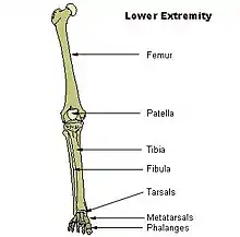
Fibular hemimelia
Fibular hemimelia or longitudinal fibular deficiency is "the congenital absence of the fibula and it is the most common congenital absence of long bone of the extremities."[1][2] It is the shortening of the fibula at birth, or the complete lack thereof. Fibula Hemimelia is a catastrophic deformity of the leg which often causes severe knee instability due to the absence of multiple ligaments including the ACL and lack of functionality in any remaining ligaments. Severe forms of Fibula Hemimelia in a patient can result in a non functional locked and malformed ankle which is significantly smaller as well as missing toes. This condition also results in a warped leg which lacks circulation and has nerve damage. In many cases of the deformity almost all types of tissue within the patients leg are deformed and seriously affected. It can lead to serious and debilitating problems and has a significant impact on a patients quality of life.[3] In humans, the disorder can be noted by ultrasound in utero to prepare for amputation after birth or complex bone lengthening surgery. The amputation usually takes place at six months with removal of portions of the legs to prepare them for prosthetic use. The other treatments which include repeated corrective osteotomies and leg-lengthening surgery (Ilizarov apparatus) are costly and associated with residual deformity.[4] It is noted however that these treatments have very little effect on the multitude of problems such as severe knee instability that fibula hemimelia causes. Fibula Hemimelia is a degenerative and lifelong condition which can result in a leg which is unable to bear any weight and can rapidly worsen without any acute injuries.[5]
| Fibular hemimelia | |
|---|---|
| Other names | Longitudinal fibular deficiency |
 | |
| Fibula Hemimelia in patient affecting right side | |
| Specialty | Medical genetics, orthopedics |
Characteristics
Characteristics are:
- A fibrous band instead of the fibula
- Short deformed leg
- Absence of the lateral part of the ankle joint (due to absence of the distal end of the fibula), and what is left is unstable; the foot has an equinovalgus deformity
- Possible absence of part of the foot requiring surgical intervention to bring the foot into normal function, or amputation.
- Possible absence of one or two toes on the foot
- Possible conjoined toes or metatarsals
Partial or total absence of fibula is among the most frequent limb anomalies. It is the most common long bone deficiency and is the most common skeletal deformity in the leg. It most often is unilateral (present only on one side). It may also present as bilateral (affecting both legs). Paraxial fibular hemimelia is the most common manifestation in which only the postaxial portion of the limb is affected. It is commonly seen as a complete terminal deficiency, where the lateral rays of the foot are also affected. Hemimelia can also be intercalary in which case the foot remains unaffected. Although the missing bone is easily identified, this condition is not simply a missing bone.[2] Males are affected twice as often as females in most series.[6]
Causes
The cause of fibular hemimelia is unclear. Purportedly, there have been some incidents of genetic distribution in a family; however, this does not account for all cases. Maternal viral infections, embryonic trauma, teratogenic environmental exposures or vascular dysgenesis (failure of the embryo to form a satisfactory blood supply) between four and seven weeks gestation are considered possible causes.[7]
In an experimental mouse model, change in the expression of a homeobox gene led to similar, but bilateral, fibular defects.[8]
Notable people
- Jessica Long – American Paralympic swimmer
- Liam Malone – New Zealand Paralympic athlete
- Aimee Mullins – American Paralympic athlete, actress, and fashion model
- Oscar Pistorius – Retired South African Paralympic and Olympic athlete and convicted murderer
- Long Jeanne Silver – American former pornographic actress
- Erik Stolhanske – American actor, writer, director, producer
See also
References
- Eze KC, Akhigbe AO, Awosanya GO (September 2007). "Fibular hemimelia: a case report". Nigerian Journal of Clinical Practice. 10 (3): 259–61. PMID 18072458.
- Achterman C, Kalamchi A (May 1979). "Congenital deficiency of the fibula". The Journal of Bone and Joint Surgery. British Volume. 61-B (2): 133–7. doi:10.1302/0301-620X.61B2.438260. PMID 438260.
- Paley D (December 2016). "Surgical reconstruction for fibular hemimelia". Journal of Children's Orthopaedics. 10 (6): 557–583. doi:10.1007/s11832-016-0790-0. PMC 5145840. PMID 27909861.
- Stanitski DF, Stanitski CL (2003). "Fibular hemimelia: a new classification system". Journal of Pediatric Orthopedics. 23 (1): 30–4. doi:10.1097/01241398-200301000-00006. PMID 12499939. S2CID 41594905.
- Alaseirlis DA, Korompilias AV, Beris AE, Soucacos PN (September 2011). "Residual malformations and leg length discrepancy after treatment of fibular hemimelia". Journal of Orthopaedic Surgery and Research. 6 (1): 51. doi:10.1186/1749-799X-6-51. PMC 3191474. PMID 21951397.
- Wheeless CR (2011-03-30). "Fibular Hemimelia: (longitudinal fibular deficiency)". Wheeless' Textbook of Orthopaedics. Wheelessonline.com. Retrieved 2012-08-03.
- "Fibular Hemimelia". orpha.net. Retrieved 2013-02-24.
- Papenbrock T, Visconti RP, Awgulewitsch A (April 2000). "Loss of fibula in mice overexpressing Hoxc11". Mechanisms of Development. 92 (2): 113–23. doi:10.1016/S0925-4773(99)00344-5. PMID 10727851. S2CID 14963600.
External links
- Journal of Joint Bone Surgery 1997 Jan;79(1):58–65.
- North American Reporting Center for Amphibian Malformations (NARCAM) Jul. 2011
- Minnesota's Malformed Frogs Jul. 2011
- Studies offer new insights into causes of deformed frogs Jul. 2011
| Classification |
|---|