Great cardiac vein
The great cardiac vein (left coronary vein) begins at the apex of the heart and ascends along the anterior longitudinal sulcus to the base of the ventricles.
| Great cardiac vein | |
|---|---|
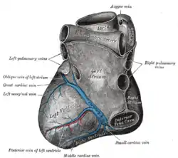 Base and diaphragmatic surface of heart. (Great cardiac vein labeled at center left.) | |
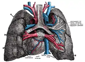 Pulmonary vessels, seen in a dorsal view of the heart and lungs. The lungs have been pulled away from the median line, and a part of the right lung has been cut away to display the air-ducts and bloodvessels (great coronary vein labeled at center bottom). | |
| Details | |
| Drains to | Coronary sinus |
| Identifiers | |
| Latin | Vena cordis magna, vena cardiaca magna |
| TA98 | A12.3.01.003 |
| TA2 | 4159 |
| FMA | 4707 |
| Anatomical terminology | |
It then curves around the left margin of the heart to reach the posterior surface. It merges with the oblique vein of the left atrium to form the coronary sinus,[1] which drains into the right atrium.
At the junction of the great cardiac vein and the coronary sinus, there is typically a valve present. This is the Vieussens valve of the coronary sinus.[1]
It receives tributaries from the left atrium and from both ventricles: one, the left marginal vein, is of considerable size, and ascends along the left margin of the heart.
References
- McAlpine, W. A. (2012). Heart and Coronary Arteries: An Anatomical Atlas for Clinical Diagnosis, Radiological Investigation, and Surgical Treatment. Springer Science & Business Media. ISBN 9783642659836.
This article incorporates text in the public domain from page 642 of the 20th edition of Gray's Anatomy (1918)
External links
- Anatomy photo:20:11-0101 at the SUNY Downstate Medical Center - "Heart: Cardiac veins"
- Anatomy figure: 20:03-05 at Human Anatomy Online, SUNY Downstate Medical Center - "Anterior view of the heart."
| Authority control |
|
|---|