Stippling (dentistry)
The gingiva often possess a textured surface that is referred to as being stippled (engraved points).[1] Stippling only presents on the attached gingiva bound to underlying alveolar bone, not the freely moveable alveolar mucosaor free gingiva . Stippling used to be thought to indicate health, but it has since been shown that smooth gingiva is not an indication of disease, unless it is smooth due to a loss of previously existing stippling.
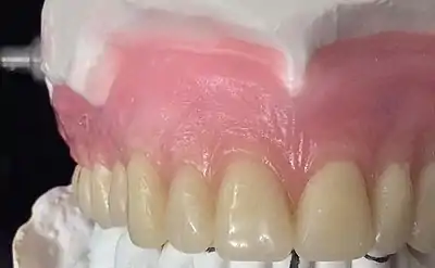
Stippling is a consequence of the microscopic elevations and depressions of the surface of the gingival tissue due to the connective tissue projections within the tissue.[1] The degree of keratinization and the prominence of stippling appear to be related.[1] To be more specific, stippling occurs at sites of fusion of the epithelial ridges (also known as rete pegs-depression of epithelium ) and correspond to the fusion of the valleys created by the connective tissue papillae(elevation of connective tissue papilla.An example of stippling could be dots found in basketball or an orange.[2]
References
- Itoiz ME, Carranza FA (2002). "The Gingiva". In Newman MG, Takei HH, Carranza FA (eds.). Carranza’s Clinical Periodontology (9th ed.). Philadelphia: W.B. Saunders Company. p. 30. ISBN 978-0-7216-8331-7.
- Lindhe J, Karring T, Lang NP. Lindhe's Clinical Periodontology and Implant Dentistry (4th ed.). ISBN 978-1-4051-0236-0.